In vitro experimental simulation study of the hemodynamics based on the FDA benchmark model
-
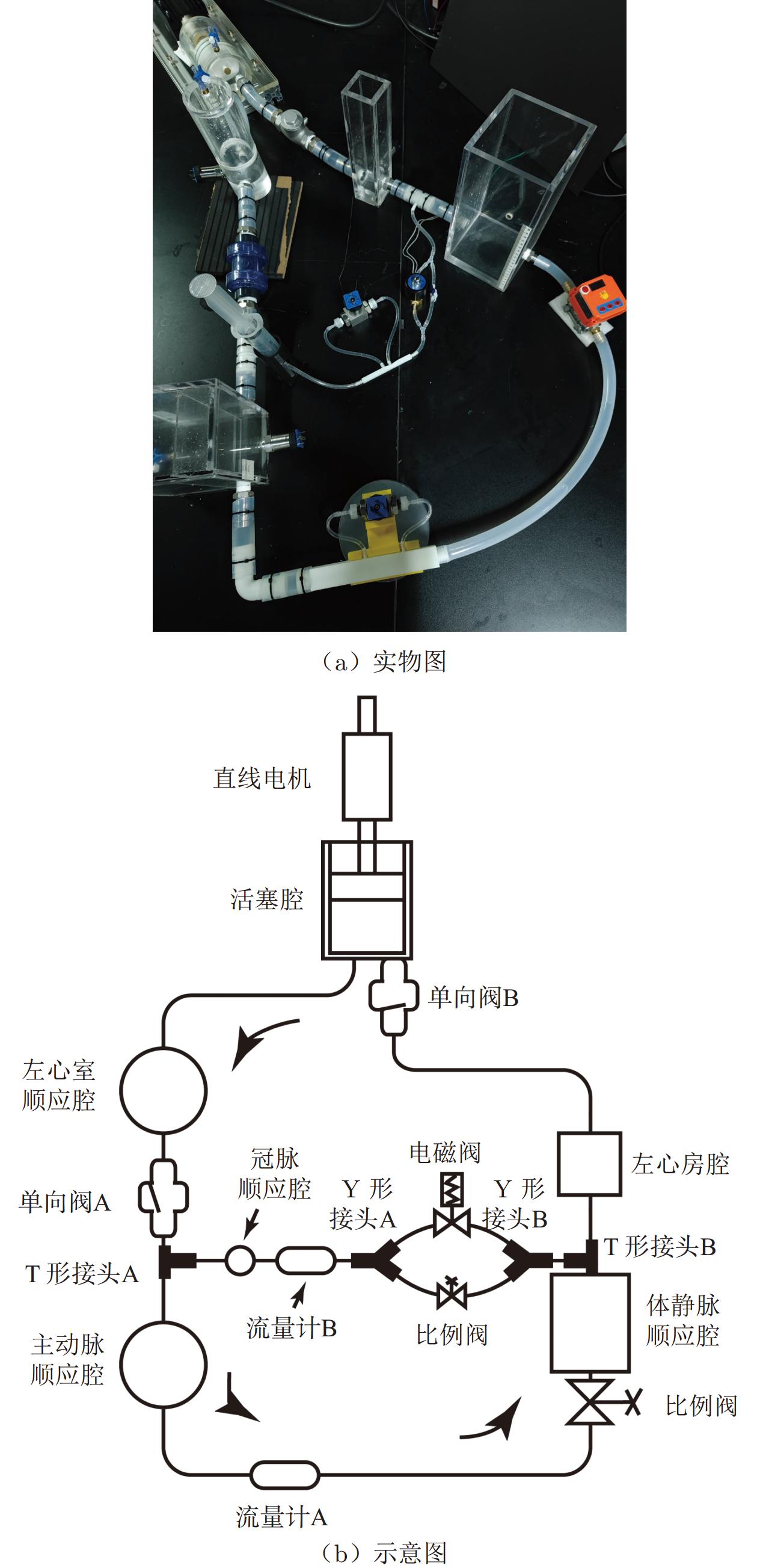
摘要: 为了离体开展冠状动脉血液动力学研究,并从流体力学角度研究心血管疾病产生机理,搭建了具有体循环和冠状动脉循环的体外模拟循环回路。以美国食品药品监督管理局(Food and Drug Administration, FDA)发布的标准喷管模型为研究对象,采用多种流体测量技术在冠状动脉流动工况下进行血液动力学离体研究:通过荧光粒子模拟血小板黏附实验,定性模拟及预测喷管模型内部血栓产生位置;采用粒子图像测速技术(Particle Image Velocimetry, PIV)测量喷管模型内部流场,定量分析血栓产生位置及对应位置血液动力学的关系。研究结果表明:荧光粒子容易在后台阶流动结构附近黏附于模型壁面,流场数据显示血栓产生位置与壁面附近的低速区和回流有关。体外血小板黏附模拟和血液动力学研究可为冠状动脉内部血栓形成、相关医疗器械研发提供参考。Abstract: In order to study the hemodynamics of the coronary artery in vitro and to understand the mechanism of cardiovascular disease from the perspective of fluid mechanics, a mock circulatory loop with systemic circulation and coronary circulation was constructed. Combined with a variety of fluid measurement techniques, the hemodynamics of the benchmark nozzle model proposed by Food and Drug Administration (FDA) was studied in vitro under the condition of coronary flow. The location of thrombus in the nozzle model was qualitatively predicted by the platelet adhesion emulation technique using fluorescent particles. The flow field inside the nozzle model was captured by particle image velocimetry (PIV), and the relationship between the thrombus formation and hemodynamics at the corresponding location was quantitatively analyzed. The results show that fluorescent particles are easy to adhere to the wall of the model near the flow structure of the backward-facing step, and the flow field data show that the thrombus formation location is related to the low velocity region and reflux near the wall. In vitro platelet adhesion emulation and hemodynamic research can provide references for coronary thrombotic investigation and related medical device development.
-
Key words:
- mock circulatory loop /
- FDA benchmark model /
- hemodynamics /
- thrombus /
- PIV
-
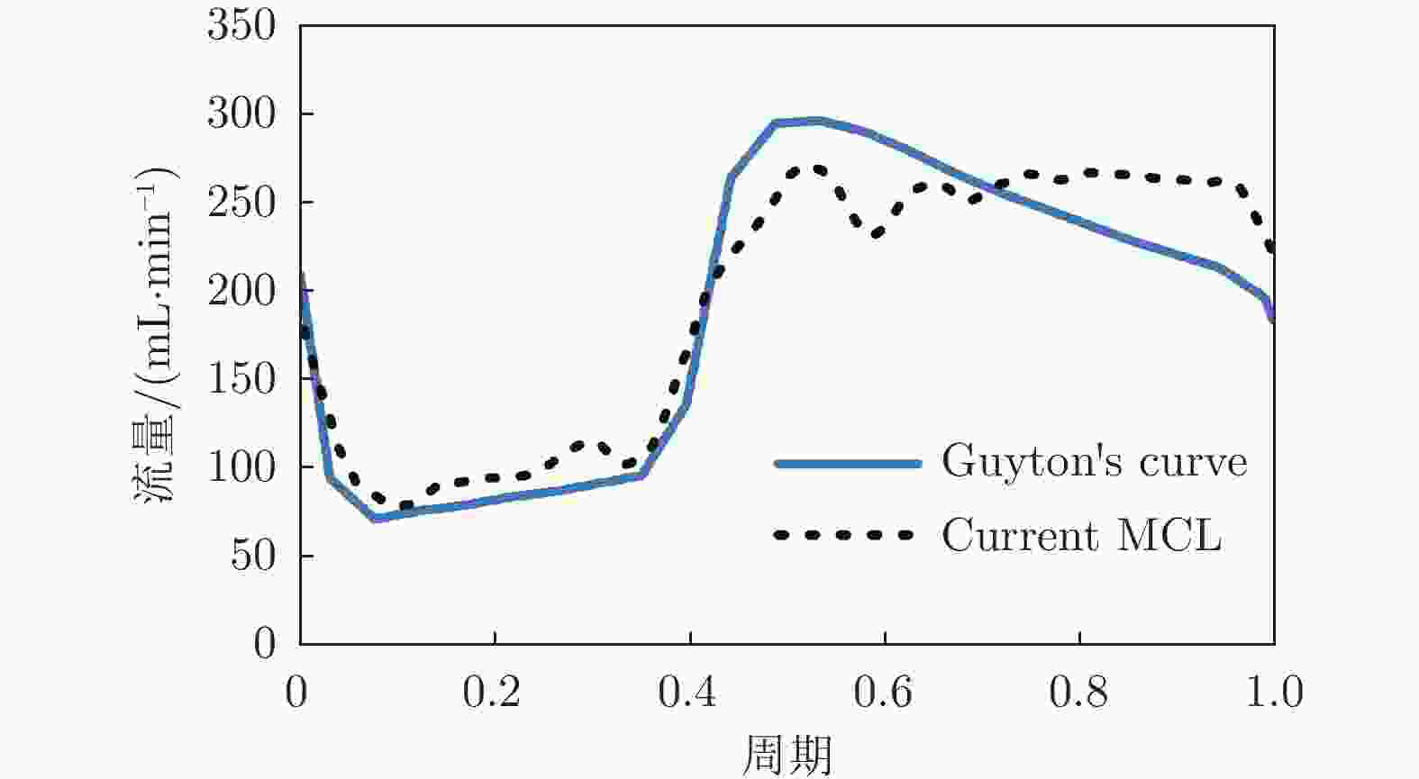
图 2 冠脉流量比较:Guyton理论曲线和本MCL平台冠脉流量曲线
Figure 2. The comparison between the coronary flow rate curve from current MCL and Guyton’s curve
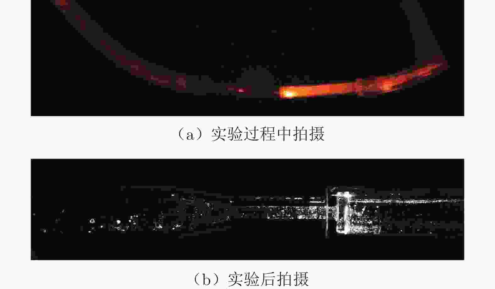
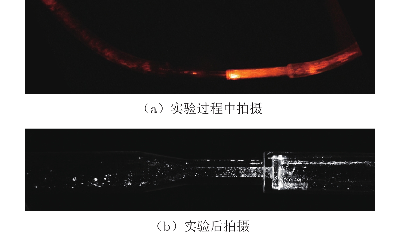
图 5 荧光粒子模拟血小板黏附实验结果
Figure 5. The results of the platelet adhesion simulation using fluorescent particles
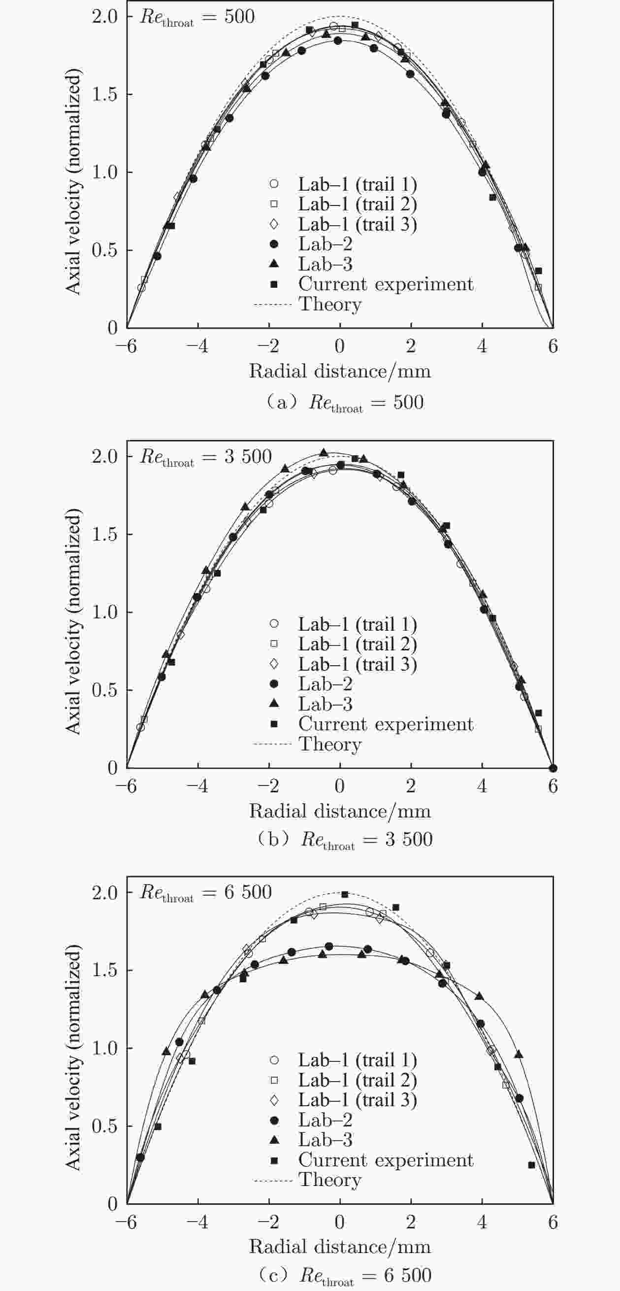
图 6 3个工况下的入口段轴向速度比较
Figure 6. Axial velocity profile comparison at the entrance section under three flow conditions
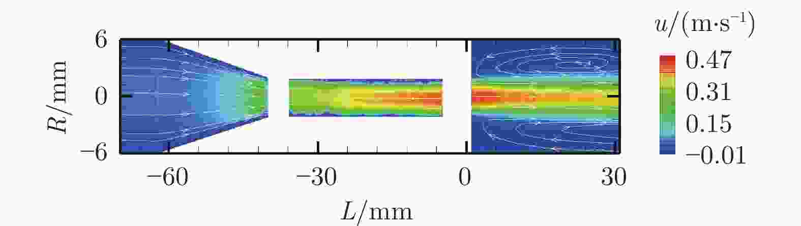
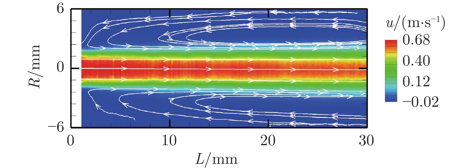
图 7 FDA标准喷管模型3个测量位置的平均轴向速度场
Figure 7. Ensemble average velocity field of three measurement locations in FDA benchmark model
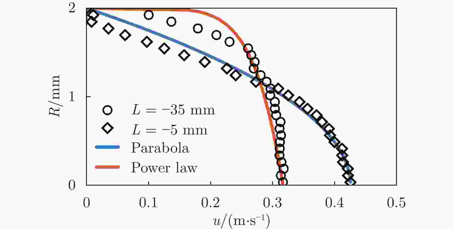
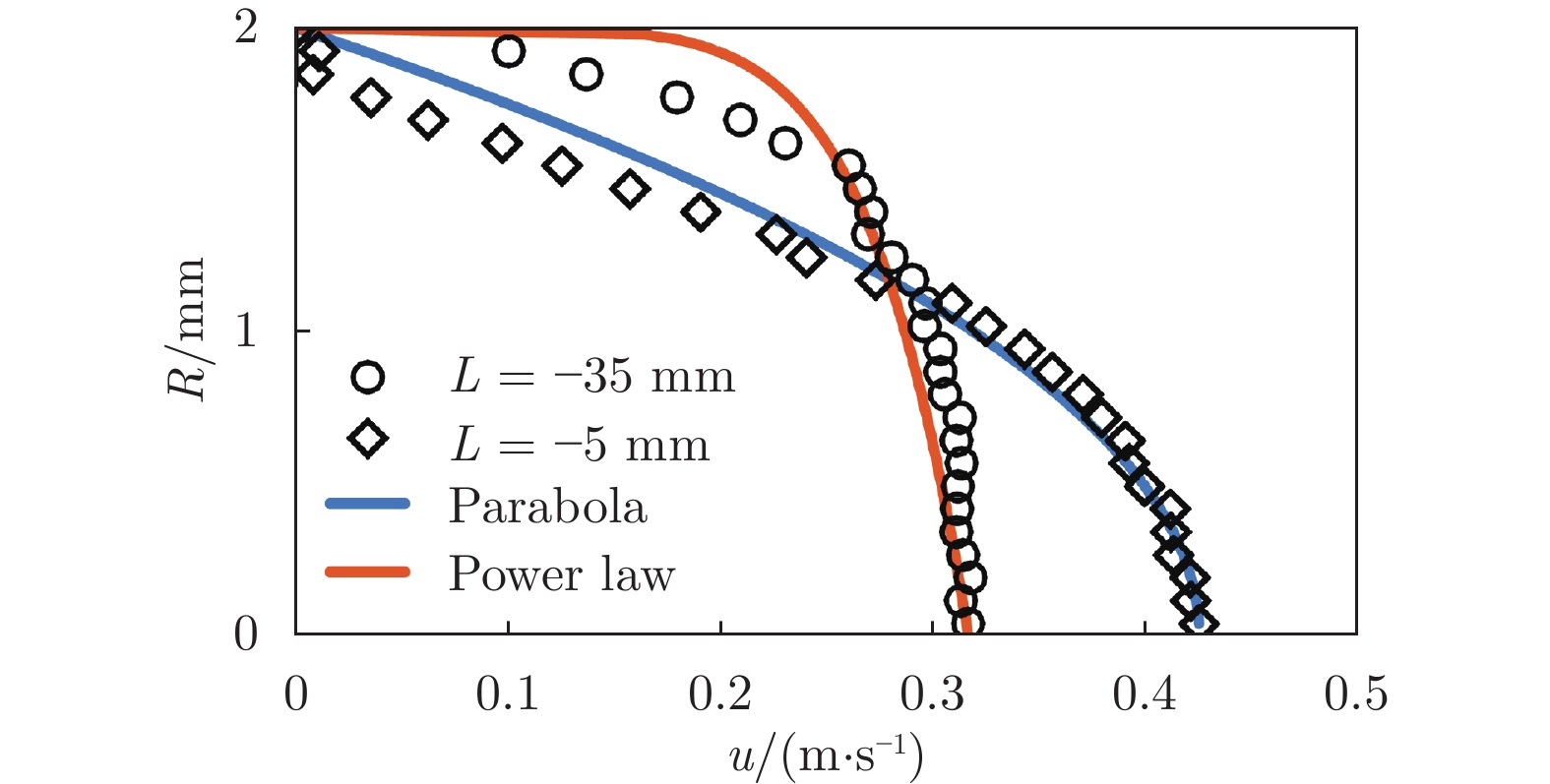
图 8 近喉部入口处(L = −35 mm)和近喉部出口处(L = −5 mm)轴向速度剖面,以及理论抛物线速度剖面和幂律函数(n = 1/8)速度剖面
Figure 8. Axial velocity profiles at the locations near inlet (L = −35 mm) and outlet (L = −5 mm) of the throat, the theoretical parabolic velocity profile and the power-law (n = 1/8) velo-city profile
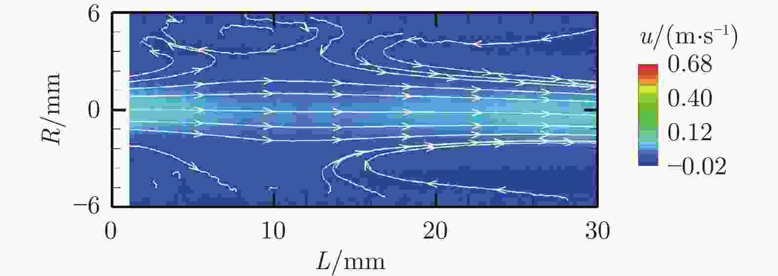
图 9 心脏收缩末期突扩段内部速度场
Figure 9. Velocity field inside the sudden expansion area at the end of systole period
-
[1] TSAO C W, ADAY A W, ALMARZOOQ Z I, et al. Heart disease and stroke statistics-2022 update: a report from the American heart association[J]. Circulation, 2022, 145(8): e153–e639. doi: 10.1161/CIR.0000000000001052 [2] ADNAN G, SINGH D P, MAHAJAN K. Coronary Artery Thrombus[M]//Treasure Island (FL): StatPearls Publishing, 2022. [3] BURKE A P, KOLODGIE F D, FARB A, et al. Healed plaque ruptures and sudden coronary death: evidence that subclinical rupture has a role in plaque progression[J]. Circulation, 2001, 103(7): 934–940. doi: 10.1161/01.cir.103.7.934 [4] AMBROSE J A, SINGH M. Pathophysiology of coronary artery disease leading to acute coronary syndromes[J]. F1000Prime Reports, 2015, 7: 08. doi: 10.12703/P7-08 [5] DAVIES M J, THOMAS A. Thrombosis and acute coronary-artery lesions in sudden cardiac ischemic death[J]. The New England Journal of Medicine, 1984, 310(18): 1137–1140. doi: 10.1056/NEJM198405033101801 [6] SALAZAH A E. Experimental myocardial infarction[J]. Circulation Research, 1961, 9(6): 1351–1356. doi: 10.1161/01.res.9.6.1351 [7] WILLERSON J T, GOLINO P, EIDT J, et al. Specific platelet mediators and unstable coronary artery lesions. Experimental evidence and potential clinical implications[J]. Circulation, 1989, 80(1): 198–205. doi: 10.1161/01.cir.80.1.198 [8] CAPPON F, WU T T, PAPAIOANNOU T, et al. Mock circulatory loops used for testing cardiac assist devices: a review of computational and experimental models[J]. The International Journal of Artificial Organs, 2021, 44(11): 793–806. doi: 10.1177/03913988211045405 [9] KHUDZARI A Z M, KADIR M R A, OSMAN K, et al. Mock circulatory loop for cardiovascular assist device testing[M]//DEWI D E O, HAU Y W, KHUDZARI A Z M, et al. Cardiovascular Engineering. Singapore: Springer, 2020: 177-200. [10] XU K W, GAO Q, WAN M, et al. Mock circulatory loop applications for testing cardiovascular assist devices and in vitro studies[J]. Frontiers in Physiology, 2023, 14: 1175919. doi: 10.3389/fphys.2023.1175919 [11] PARK Y K, MITA Y, OKI E, et al. Quantitative Evaluation for Anastomotic Technique of Coronary Artery Bypass Grafting by using In-vitro Mock Circulatory System[C]//Proc of the 2007 29th Annual International Conference of the IEEE Engineering in Medicine and Biology Society. 2007: 2705-2708. [12] PARK Y K, MITA Y, OKI E, et al. Development of “Patient Robot”; Training Robot based on Quantitative Analysis of Surgical Technique[C]//Proc of the First IEEE/RAS-EMBS International Conference on Biomedical Robotics and Biomechatronics. 2006: 318-322. [13] CALDERAN J, MAO W B, SIROIS E, et al. Development of an in vitro model to characterize the effects of transcatheter aortic valve on coronary artery flow[J]. Artificial Organs, 2016, 40(6): 612–619. doi: 10.1111/aor.12589 [14] STOCK S, SCHARFSCHWERDT M, MEYER-SARAEI R, et al. In vitro coronary flow after transcatheter aortic valve-in-valve implantation: a comparison of 2 valves[J]. The Journal of Thoracic and Cardiovascular Surgery, 2017, 153(2): 255-263. E1. [15] HORNY L, CHLUP H, VESELY J, et al. In vitro coronary stent implantation: vessel wall-stent interaction[C]//JOBBÁGY Á. 5th European Conference of the International Federation for Medical and Biological Engineering: Vol. 37. Berlin, Heidelberg: Springer, 2011: 795-798. [16] HARIHARAN P, GIARRA M, REDDY V, et al. Multilaboratory particle image velocimetry analysis of the FDA benchmark nozzle model to support validation of computational fluid dynamics simulations[J]. Journal of Biomechanical Engineering, 2011, 133(4): 041002. doi: 10.1115/1.4003440 [17] RABEN J S, HARIHARAN P, ROBINSON R, et al. Time-resolved particle image velocimetry measurements with wall shear stress and uncertainty quantification for the FDA nozzle model[J]. Cardiovascular Engineering and Technology, 2016, 7(1): 7–22. doi: 10.1007/s13239-015-0251-9 [18] STEWART S F C, PATERSON E G, BURGREEN G W, et al. Assessment of CFD performance in simulations of an idealized medical device: results of FDA’s first computational interlaboratory study[J]. Cardiovascular Engineering and Technology, 2012, 3(2): 139–160. doi: 10.1007/s13239-012-0087-5 [19] STIEHM M, WÜSTENHAGEN C, SIEWERT S, et al. Numerical simulation of pulsatile flow through a coronary nozzle model based on FDA’s benchmark geometry[J]. Current Directions in Biomedical Engineering, 2017, 3(2): 775–778. doi: 10.1515/cdbme-2017-0163 [20] GUYTON A C, HALL J E. Textbook of medical physiology[M]. 11th ed. Philadelphia: Elsevier Saunders, 2006. [21] LIU G M, ZHANG Y, CHEN H B, et al. Platelet deposition estimation: a novel method for emulating the pump thrombosis potential of blood pumps[J]. Artificial Organs, 2020, 44(5): 465–472. doi: 10.1111/aor.13620 [22] LIU G M, CHEN H B, HOU J F, et al. Platelet adhesion emulation: a novel method for estimating the device thrombosis potential of a ventricular assist device[J]. The International Journal of Artificial Organs, 2020, 43(4): 252–257. doi: 10.1177/0391398819885946 -

 下载:
下载:


 点击查看大图
计量
- 文章访问数: 28
- HTML全文浏览量: 7
- PDF下载量: 1
- 被引次数: 0